
购物车
 您的购物车当前为空

 很棒
很棒Neratinib (HKI-272) 是一种酪氨酸激酶受体抑制剂,可以抑制 HER2 和 EGFR (IC50=59/92 nM),具有不可逆性和口服活性。Neratinib 具有抗肿瘤活性,可以用于治疗乳腺癌。
很棒纯度: 99.85%
别名 来那替尼, HKI-272
Neratinib (HKI-272) 是一种酪氨酸激酶受体抑制剂,可以抑制 HER2 和 EGFR (IC50=59/92 nM),具有不可逆性和口服活性。Neratinib 具有抗肿瘤活性,可以用于治疗乳腺癌。
| 规格 | 价格 | 库存 | 数量 |
|---|---|---|---|
| 5 mg | ¥ 282 | 现货 | |
| 10 mg | ¥ 497 | 现货 | |
| 25 mg | ¥ 788 | 现货 | |
| 50 mg | ¥ 1,080 | 现货 | |
| 100 mg | ¥ 1,890 | 现货 | |
| 200 mg | ¥ 3,490 | 现货 | |
| 500 mg | ¥ 5,580 | 现货 |
TargetMol的所有产品仅用作科学研究或药证申报,不能被用于人体,我们不向个人提供产品和服务。请您遵守承诺用途,不得违反法律法规规定用于任何其他用途。
凭借在化合物合成方面的丰富经验,我们可以根据您的研究需求为该产品提供快速定制合成服务。
| 产品描述 | Neratinib (HKI-272) is a tyrosine kinase receptor inhibitor that inhibits HER2 and EGFR (IC50=59/92 nM) with irreversible and oral activity. Neratinib has antitumor activity and can be used to treat breast cancer. |
| 靶点活性 | HER2:59 nM (cell free), EGFR:92 nM (cell free) |
| 体外活性 | 方法:36 种乳腺癌细胞系用 Neratinib 处理 5 天,使用 cell counting and acid phosphatase assay 检测细胞增殖。 |
| 体内活性 | 方法:为检测体内抗肿瘤活性,将 Neratinib (20-80 mg/kg in 0.5% methocellulose-0.4% Tween 80) 口服给药给携带 3T3/neu 异种移植瘤的 nude 小鼠,每天一次,持续十天。 |
| 激酶实验 | Activity of HER-2 and EGFR cytoplasmic domains was measured by an autophosphorylation assay using time-resolved fluorometry. Compounds were prepared as 10 mg/ml stocks in DMSO and diluted in 25 mm HEPES (pH 7.5; 0.002 ng/ml–20 μg/ml). Enzyme [diluted in 100 mm HEPES (pH 7.5) and 50% glycerol] was incubated with inhibitor in 4 mm HEPES (pH 7.5), 0.4 mm MnCl2, 20 μm sodium vanadate, and 0.2 mm DTT for 15 min at room temperature in 96-well ELISA plates. The kinase reaction was initiated by the addition of 40 μm ATP and 20 mm MgCl2 and allowed to proceed for 1 h at room temperature. Plates were washed, and phosphorylation was detected using Europium-labeled anti-phospho-tyrosine antibodies (15 ng/well). After washing and enhancement steps according to the manufacturer's recommendations, signal was detected using a Victor^2 fluorescence reader (excitation wavelength 340 nm, emission wavelength 615 nm). The concentration of compound that inhibited receptor phosphorylation by 50% (IC50) was calculated from inhibition curves [1]. |
| 细胞实验 | Cells were plated in 96-well tissue culture plates (3T3, 3T3/neu, 5000 cells/well; A431, SK-Br-3, BT474, MDA-MB-435, and SW620, 10,000 cells/well). The following day, dilutions of compound (0.5 ng/ml–5 μg/ml) were added, and cells were cultured for 2 days (6 days for BT474). Cell proliferation was determined using sulforhodamine B, a protein binding dye. Briefly, cells were fixed with 10% trichloroacetic acid and washed extensively with water. Cells were then stained with 0.1% sulforhodamine B and washed in 5% acetic acid. Protein-associated dye was solubilized in 10 mm Tris, and absorbance was measured at 450 nm (Victor^2). Inhibition of cell proliferation was calculated using the formula: percentage of inhibition = 100 ? 100 (Td ? To/Tc ? To), where Td is the absorbance of drug-treated cells, Tc is the absorbance of untreated cells, and To is the absorbance at the time of drug addition. To values were determined by plating cells separately and fixing them at the time of drug addition. The concentration of compound which inhibits cell proliferation by 50% (IC50) was determined from inhibition curves [1]. |
| 动物实验 | Athymic female nude mice (5 animals/group) were implanted s.c. with BT474 tumor fragments (~30 mm^3). When tumors reached 200–300 mg, animals were given a single oral dose (40 mg/kg) of HKI-272 in pH 2.0 water. Tumors from control and treated animals were excised at 1, 3, 6, and 24 h and minced. Tumor fragments were suspended in 10 mm Tris (pH 7.5), 5 mm EDTA, 150 mm NaCl, 1% Triton X-100, 1% sodium deoxycholate, 0.1% SDS, 1 mm phenylmethylsulfonyl fluoride, 10 μg/ml pepstatin, 10 μg/ml leupeptin, 10 μg/ml aprotinin, 2 mm sodium vanadate, and 100 mm sodium fluoride and lysed by homogenization on ice with a polytron. After clarification by centrifugation, protein concentration in lysates was estimated using the Bio-Rad DC protein assay. Sixty μg of lysate pooled from each group were analyzed by SDS-PAGE and immunoblotting with phospho-tyrosine-specific antibodies. Pooled extracts were also immunoprecipitated using 4 μg of anti-HER-2 antibodies for 1 h at 4°C. Immune complexes were collected on protein A-agarose, washed, and analyzed by immunoblotting using phospho-tyrosine-specific antibodies. Extracts from individual tumors were analyzed to determine variability between animals [1]. |
| 别名 | 来那替尼, HKI-272 |
| 分子量 | 557.04 |
| 分子式 | C30H29ClN6O3 |
| CAS No. | 698387-09-6 |
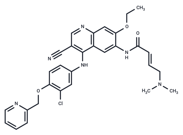
| Smiles | N(C=1C2=C(C=C(OCC)C(NC(/C=C/CN(C)C)=O)=C2)N=CC1C#N)C3=CC(Cl)=C(OCC4=CC=CC=N4)C=C3 |
| 密度 | 1.337 g/cm3 |
| 存储 | Powder: -20°C for 3 years | In solvent: -80°C for 1 year Shipping with blue ice/Shipping at ambient temperature. 实际储存温度请以COA为准 | |||||||||||||||
| 溶解度信息 | H2O: < 1 mg/mL (insoluble or slightly soluble) Ethanol: < 1 mg/mL (insoluble or slightly soluble) DMSO: 5 mg/mL (8.98 mM), Sonication is recommended. | |||||||||||||||
| 体内实验配方 | 10% DMSO+40% PEG300+5% Tween 80+45% Saline: 0.25 mg/mL (0.45 mM), Solution. 请按顺序添加溶剂,在添加下一种溶剂之前,尽可能使溶液澄清。如有必要,可通过加热、超声、涡旋处理进行溶解。工作液建议现配现用。以上配方仅供参考,体内配方并不是绝对的,请根据不同情况进行调整。 | |||||||||||||||
溶液配制表 | ||||||||||||||||
DMSO
该溶液配制表仅适用于固体产品。对于液体产品,请根据标明的浓度或密度计算稀释方案。 | ||||||||||||||||
对于不同动物的给药剂量换算,您也可以参考 更多
 嗨!有任何问题?点我咨询
版权所有©2015-2026 TargetMol Chemicals Inc.保留所有权利.
沪ICP备20019793号-4 | 沪公网安备 31010602006700号 | 沪(静)应急管危经许[2024]203441