
购物车
 您的购物车当前为空

 很棒
很棒纯度: 99.81%
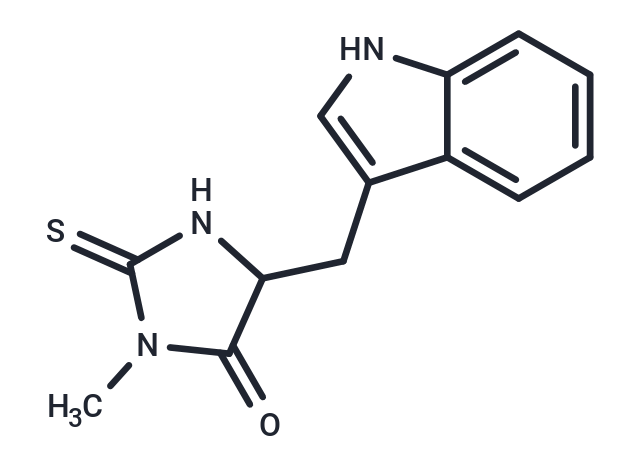
别名 Necrostatin 1, Nec-1
Necrostatin-1 (Nec-1) 是一种坏死性凋亡抑制剂和 RIP1 抑制剂,具有特异性。Necrostatin-1 抑制 TNF-α 诱导的坏死性凋亡。Necrostatin-1 也可以抑制 IDO。

为众多的药物研发团队赋能,
让新药发现更简单!
很棒纯度: 99.81%
Necrostatin-1 (Nec-1) 是一种坏死性凋亡抑制剂和 RIP1 抑制剂,具有特异性。Necrostatin-1 抑制 TNF-α 诱导的坏死性凋亡。Necrostatin-1 也可以抑制 IDO。
| 规格 | 价格 | 库存 | 数量 |
|---|---|---|---|
| 1 mg | ¥ 167 | 现货 | |
| 5 mg | ¥ 367 | 现货 | |
| 10 mg | ¥ 589 | 现货 | |
| 25 mg | ¥ 898 | 现货 | |
| 50 mg | ¥ 1,230 | 现货 | |
| 100 mg | ¥ 1,630 | 现货 | |
| 200 mg | ¥ 2,450 | 现货 | |
| 500 mg | ¥ 3,970 | 5日内发货 | |
| 1 mL x 10 mM (in DMSO) | ¥ 423 | 现货 |
| 产品描述 | Necrostatin-1 (Nec-1) is a necrotic apoptosis inhibitor and RIP1 inhibitor with specificity. Necrostatin-1 inhibits TNF-α-induced necrotic apoptosis. Necrostatin-1 also inhibits IDO. |
| 靶点活性 | Jurkat cells:494 nM, Sf9 cells:182 nM, RIP1:490 nM (EC50, Jurkat cells), RIP1:182 nM (EC50) |
| 体外活性 | 方法:人肝癌细胞 Huh7 和 SK-HEP-1 用 Necrostatin-1 (10-20 µM) 预处理 1 h,再用 sulfasalazine、 erastin 或 RSL3 处理24 h,使用 CellTiter Glo® assay 检测细胞活力。 |
| 体内活性 | 方法:为研究造影剂诱导的 AKI (CIAKI) 的病理生理学,将 Necrostatin-1 (1.65 mg/kg) 单次腹腔注射给 C57BL/6 小鼠,15 min 后使用放射性造影剂 (RCM) 诱导 CIAKI。 |
| 激酶实验 | The assay was performed essentially as described. 293T cells were transfected with pcDNA3-FLAG-RIP1 vector, vectors encoding RIP1 mutant proteins or pcDNA3-RIP2-Myc and pcDNA3-FLAG-RIP3 vectors using standard Ca3(PO4)2 precipitation procedure. Culture medium was replaced 6 h after the transfection and cells were lysed 48 h later in the TL buffer consisting of 1% Triton X-100, 150 mM NaCI, 20 mM HEPES, pH 7.3, 5 mM EDTA, 5 mM NaF, 0.2 mM NaVO3 and complete protease inhibitor cocktail. Immunoprecipitation was carried out for 16 h at 4 °C using anti-FLAG M2 agarose beads, followed by three washes with TL buffer and two washes with 20 mM HEPES, pH 7.3. Beads were incubated in 15 μl of the reaction buffer containing 20 mM HEPES, pH 7.3, 10 mM MnCl2 and 10 mM MgCl2 for 15 min at 23–25 °C in the presence of different concentrations of necrostatins. For these assays, compound stocks (in DMSO) were diluted to appropriate concentrations in DMSO before the addition to the reactions to maintain final concentration of DMSO for all samples at 3%. Kinase reaction was initiated by addition of 10 μM cold ATP and 1 mCi of [γ-32P] ATP, and reactions were carried out for 30 min at 30 °C. Reactions were stopped by boiling in SDS-PAGE sample buffer and subjected to 8% SDS-PAGE. RIP1 band was visualized by analysis in a Storm 8200 Phosphorimager. Similar protocol was used for endogenous RIP1 kinase reactions, except mouse monoclonal RIP1 antibody and protein magnetic beads or rabbit RIP1 antibody-coupled agarose beads were used. For recombinant baculovirally expressed RIP1, protein was expressed in Sf9 cells according to manufacturer's instructions and purified using glutathione-sepharose beads. Protein was eluted in 50 mM Tris-HCl, pH 8.0 supplemented with 10 mM reduced glutathione, and eluted protein was used in the kinase reactions, supplemented with 5 × kinase reaction buffer (100 mM HEPES, pH 7.3, 50 mM MnCl2, 50 mM MgCl2, 50 μM cold ATP and 5 μCi of [γ-32P]ATP) [1]. |
| 细胞实验 | Determination of EC50 was performed in FADD-deficient Jurkat cells treated with human TNFα as previously described. Briefly, cells were seeded into 96-well plates and treated with a range of necrostatin concentrations (30 nM to 100 μM, 11 dose points) in the presence and absence of 10 ng ml–1 human TNFα for 24 h. For these and all other cellular assays, compound stocks (in DMSO) were diluted to appropriate concentrations in DMSO before addition to the cells to maintain final concentration of DMSO for all samples at 0.5%. Cell viability was determined using CellTiter-Glo luminescent cell viability assay. Ratio of luminescence in compound and TNF-treated wells to compound-treated, TNF-untreated wells was calculated (viability, %) [1]. |
| 动物实验 | 24 hours after reperfusion, mice received intravenous application of 200 μl PBS or RCM via the tail vein. A single dose of zVAD (10 mg/kg body weight) or Nec-1 (1.65 mg/kg body weight) was applied intraperitoneally 15 min. before RCM-injection. To test the activity of zVAD, we applied zVAD from the same byculture to anti-Fas-treated Jurkat cells to assure its quality before mice were treated with this compound. Mice were harvested another 24 hours after RCM-application (48 hours after reperfusion). Blood samples were obtained from retroorbital bleeding and serum levels of urea and creatinine 5 were determined according to clinical standards in the central laboratory of the University Hospital Schleswig-Holstein, Campus Kiel, Germany, employing an enzymatic ultraviolettest for urea and an enzymatic peroxidase-dependent test for creatinine according to the manufacturer's instructions. Kidneys were conserved for histology. In addition to the demonstrated experiments, we compared the PBS group to mice that only received IRI without 200 μl of PBS and detected no changes in serum concentrations of urea and creatinine or histologically [3]. |
| 别名 | Necrostatin 1, Nec-1 |
| 分子量 | 259.33 |
| 分子式 | C13H13N3OS |
| CAS No. | 4311-88-0 |
| Smiles | CN1C(=S)NC(Cc2c[nH]c3ccccc23)C1=O |
| 密度 | 1.40 g/cm3 at 20℃ |
| 存储 | Powder: -20°C for 3 years | In solvent: -80°C for 1 year | Shipping with blue ice/Shipping at ambient temperature. | |||||||||||||||||||||||||||||||||||
| 溶解度信息 | DMSO: 245 mg/mL (944.74 mM), Sonication is recommended. | |||||||||||||||||||||||||||||||||||
| 体内实验配方 | 10% DMSO+40% PEG300+5% Tween 80+45% Saline: 4 mg/mL (15.42 mM), Solution. 请按顺序添加溶剂,在添加下一种溶剂之前,尽可能使溶液澄清。如有必要,可通过加热、超声、涡旋处理进行溶解。工作液建议现配现用。以上配方仅供参考,体内配方并不是绝对的,请根据不同情况进行调整。 | |||||||||||||||||||||||||||||||||||
溶液配制表 | ||||||||||||||||||||||||||||||||||||
DMSO
| ||||||||||||||||||||||||||||||||||||
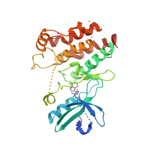
Crystal structure of RIP1 kinase in complex with necrostatin-1 analog
对于不同动物的给药剂量换算,您也可以参考 更多
 嗨!有任何问题?点我咨询
版权所有©2015-2026 TargetMol Chemicals Inc.保留所有权利.
沪ICP备20019793号-4 | 沪公网安备 31010602006700号 | 沪(静)应急管危经许[2024]203441