 您的购物车当前为空
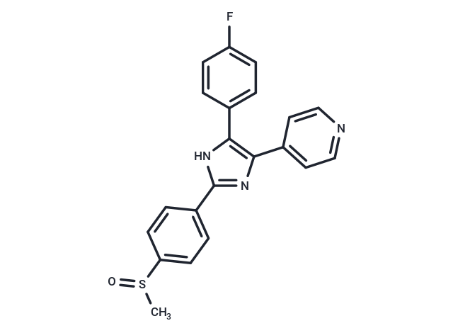
Adezmapimod
一键复制产品信息别名 SB203580, RWJ 64809, PB 203580, 4-(4-氟苯基)-2-(4-甲基亚磺酰基苯基)-5-(4-吡啶基)-1H-咪唑
Adezmapimod (SB 203580) 是一种 p38 MAPK 抑制剂 (IC50=0.3-0.5 μM),具有选择性和 ATP 竞争性。Adezmapimod 具有自噬和线粒体自噬的激活活性。Adezmapimod 显示出比 PKB、LCK 和 GSK-3β 高 100 倍以上的选择性。


为众多的药物研发团队赋能,
让新药发现更简单!
Adezmapimod
一键复制产品信息Adezmapimod (SB 203580) 是一种 p38 MAPK 抑制剂 (IC50=0.3-0.5 μM),具有选择性和 ATP 竞争性。Adezmapimod 具有自噬和线粒体自噬的激活活性。Adezmapimod 显示出比 PKB、LCK 和 GSK-3β 高 100 倍以上的选择性。
| 规格 | 价格 | 库存 | 数量 |
|---|---|---|---|
| 5 mg | ¥ 297 | 现货 | |
| 10 mg | ¥ 488 | 现货 | |
| 25 mg | ¥ 783 | 现货 | |
| 50 mg | ¥ 1,160 | 现货 | |
| 100 mg | ¥ 1,730 | 现货 | |
| 200 mg | ¥ 2,580 | 现货 | |
| 500 mg | ¥ 4,270 | 现货 | |
| 1 mL x 10 mM (in DMSO) | ¥ 348 | 现货 |
产品介绍
| 产品描述 | Adezmapimod (SB 203580) is a p38 MAPK inhibitor (IC50=0.3-0.5 μM) that is selective and ATP-competitive. Adezmapimod possesses autophagy and mitochondrial autophagy activating activity. Adezmapimod displays more than 100-fold higher selectivity than PKB, LCK, and GSK-3β. |
| 靶点活性 | p38β2:500 nM, p38:50 nM, PKB:3-5 μM (THP-1 cells), p38 MAPK:0.3-0.5 μM (THP-1 cells) |
| 体外活性 | 方法:人肝癌细胞 HepG2 用 Adezmapimod (0.1-20 μM) 处理 30 min,使用 Western Blot 方法检测靶点蛋白表达水平。 |
| 体内活性 | 方法:为检测体内活性,将 Adezmapimod (25 mg/kg in 4% DMSO+30% PEG 300+5% Tween 80+61% ddH2O) 单次腹腔注射给 LPS 处理的 C57BL/6J 小鼠,24 和 72 h 后,处死小鼠。 |
| 激酶实验 | Cells were lysed in Buffer A for Western blotting and PKB kinase assays. Kinase assays were performed according to the manufacturer's instructions. Briefly, 4 μg of sheep anti-PKBα was immobilized on 25 μl of protein G-Sepharose overnight (or 1.5 h) and washed in Buffer A (50 mM Tris, pH 7.5, 1 mM EDTA, 1 mMEGTA, 0.5 mM Na3VO4, 0.1% β-mercaptoethanol, 1% Triton X-100, 50 mM sodium fluoride, 5 mM sodium pyrophosphate, 0.1 mM phenylmethylsulfonyl fluoride, 1 μg/ml aprotinin, pepstatin, leupeptin, and 1 μM microcystin). The immobilized anti-PKB was then incubated with 0.5 ml of lysate (from 5 × 10^6 cells) for 1.5 h and washed three times in 0.5 ml of Buffer A supplemented with 0.5 M NaCl, two times in 0.5 ml of Buffer B (50 mM Tris-HCl, pH 7.5, 0.03% (w/v) Brij-35, 0.1 mM EGTA, and 0.1% β-mercaptoethanol), and twice with 100 μl of assay dilution buffer; 5× assay dilution buffer is 100 mM MOPS, pH 7.2, 125 mMβ-glycerophosphate, 25 mM EGTA, 5 mM sodium orthovanadate, 5 mM DTT. To the PKB enzyme immune complex was added 10 μl of assay dilution buffer, 40 μM protein kinase A inhibitor peptide, 100 μM PKB-specific substrate peptide, and 10 μCi of [γ-32P]ATP, all made up in assay dilution buffer. The reaction was incubated for 20 min at room temperature with shaking, then samples were pulse spun, and 40 μl of reaction volume were removed into another tube to which was added 20 μl of 40% trichloroacetic acid to stop the reaction. This was mixed and incubated for 5 min at room temperature, and 40 μl was transferred onto P81 phosphocellulose paper and allowed to bind for 30 s. The P81 pieces were washed three times in 0.75% phosphoric acid then in acetone at room temperature. γ-32P incorporation was then measured by scintillation counting [1]. |
| 细胞实验 | The luciferase reporter plasmid pIL6luc(-122) and the CAT reporter plasmid p(TRE)5CAT were transfected into TF-1 cell line by means of electroporation. Prior to transfection, cells were cultured for 16?h at a density of 0.5×10^6 cells/ml in the appropriate medium, washed twice and resuspended in RPMI 1640 at a density of 10×10^6 in 200?μl. When transfected with a single plasmid, 25?μg of DNA was added and the mixture was left at room temperature for 15?min. Cotransfections were performed with 15?μg of the reporter plasmid pIL6luc(-122) together with 15?μg of the dominant-negative expression plasmids (pRSV-MKK3(Ala), pcDNA3-MKK6(K82A), pRSV-NΔRaf1, pcDNA3-MKK4(Ala), pcDNA3-Flag-JNK1, or pcDNA3 (empty vector). Cotransfections of pGAL4tkluc (5?μg) with either pGAL4p65 (5?μg) or pGAL4dbd (5?μg) were performed under similar conditions. In addition, cells were cotransfected with 2?μg of a CMV-CAT plasmid, to normalize for transfection efficiency. Electroporation, in 0.4?cm electroporation cuvettes, was performed at 240?V and 960?μF with Gene Pulser electroporator. After electroporation, the cells were replated in RPMI 1640 containing 2% FBS. Six hours after transfection cells were stimulated for 24?h with medium or OA (30?ng/ml) or SB203580 for 30?min prior to OA stimulation. The cells were then harvested and lysed by commercially available luciferase lysis buffer. One-hundred μl of lysis product was added to 100?μl of luciferase assay reagents and luciferase activity was measured with the Anthos Lucy1 luminometer. CAT reporter activity of 100?μl lysis product plus 100?μl CAT dilution buffer was determined with a commercially available CAT Elisa kit [3]. |
| 动物实验 | In survival studies, C57BL/6J mice weighing 20 g to 30 g were briefly anesthetized with isoflurane and challenged with 0.05 mL of IT normal saline (NS, noninfected controls) or E. coli (15 × 10^9 CFU/kg) as previously described. One hour before NS challenge, mice (n = 24) received either intraperitoneal SB203580 (100 mg/kg in 0.25 mL) or diluent only (placebo). Infected animals received SB203580 in doses of 100, 10, 1, or 0.1 mg/kg or placebo 1 hour before IT E. coli (n = 241); SB203580 100 or 0.1 mg/kg or placebo 1 hour after E. coli (n = 121); or SB203580 100 mg/kg or placebo 12 hours after E. coli (n = 72). All animals received ceftriaxone (100 mg/kg in 0.1 mL, subcutaneously) for 4 days and NS (0.5 mL, subcutaneously) for 1 day beginning 4 hours after challenge. Animals were observed every 2 hours for the initial 48 hours, every 4 hours from 48 hours to 72 hours, every 8 hours from 72 hours to 96 hours, and then twice daily until study completion (168 hours). Sequential weekly experiments with 24 animals each compared either two to three doses of SB203580 versus placebo administered at similar times or similar doses of SB203580 versus placebo at differing treatment times. Study groups in each experiment were of equivalent sample size (i.e., 6 – 8 per group) [5]. |
| 别名 | SB203580, RWJ 64809, PB 203580, 4-(4-氟苯基)-2-(4-甲基亚磺酰基苯基)-5-(4-吡啶基)-1H-咪唑 |
| 分子量 | 377.43 |
| 分子式 | C21H16FN3OS |
| CAS No. | 152121-47-6 |
| Smiles | FC1=CC=C(C2=C(N=C(N2)C3=CC=C(S(C)=O)C=C3)C=4C=CN=CC4)C=C1 |
| 密度 | 1.42 g/cm3 |
| 存储 | Powder: -20°C for 3 years | In solvent: -80°C for 1 year | Shipping with blue ice/Shipping at ambient temperature. | |||||||||||||||||||||||||||||||||||
| 溶解度信息 | DMSO: 101 mg/mL (267.6 mM), Sonication is recommended. | |||||||||||||||||||||||||||||||||||
| 体内实验配方 | 10% DMSO+40% PEG300+5% Tween 80+45% Saline: 5 mg/mL (13.25 mM), Solution. 请按顺序添加溶剂,在添加下一种溶剂之前,尽可能使溶液澄清。如有必要,可通过加热、超声、涡旋处理进行溶解。工作液建议现配现用。以上配方仅供参考,体内配方并不是绝对的,请根据不同情况进行调整。 | |||||||||||||||||||||||||||||||||||
溶液配制表 | ||||||||||||||||||||||||||||||||||||
DMSO
| ||||||||||||||||||||||||||||||||||||
计算器
体内实验配液计算器
以上为“体内实验配液计算器”的使用方法举例,并不是具体某个化合物的推荐配制方式,请根据您的实验动物和给药方式选择适当的溶解方案。
剂量转换
对于不同动物的给药剂量换算,您也可以参考 更多


 很棒
很棒
 |
|