购物车
- 全部删除
 您的购物车当前为空

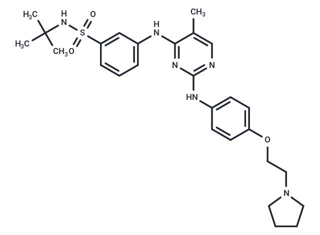
Fedratinib (TG-101348) 是一种 JAK2 抑制剂,具有选择性、ATP 竞争性和具有口服活性,抑制 JAK2 和 JAK2V617F 激酶 (IC50=3 nM)。Fedratinib 可诱导肿瘤细胞凋亡,并可用于骨髓增生性疾病的研究。

Fedratinib (TG-101348) 是一种 JAK2 抑制剂,具有选择性、ATP 竞争性和具有口服活性,抑制 JAK2 和 JAK2V617F 激酶 (IC50=3 nM)。Fedratinib 可诱导肿瘤细胞凋亡,并可用于骨髓增生性疾病的研究。
| 规格 | 价格 | 库存 | 数量 |
|---|---|---|---|
| 2 mg | ¥ 265 | 现货 | |
| 5 mg | ¥ 379 | 现货 | |
| 10 mg | ¥ 543 | 现货 | |
| 25 mg | ¥ 773 | 现货 | |
| 100 mg | ¥ 1,198 | 现货 | |
| 200 mg | ¥ 1,995 | 现货 | |
| 500 mg | ¥ 4,280 | 现货 | |
| 1 mL x 10 mM (in DMSO) | ¥ 481 | 现货 |
| 产品描述 | Fedratinib (TG-101348) is a JAK2 inhibitor that is selective, ATP-competitive, and orally active, inhibiting JAK2 and JAK2V617F kinases (IC50=3 nM). Fedratinib induces apoptosis in tumor cells and may be used in myeloproliferative diseases. |
| 靶点活性 | JAK2:3 nM (cell free), FLT3:15 nM (cell free), JAK2 (V617F):3 nM (cell free), RET:48 nM (cell free) |
| 体外活性 | 方法:人成红细胞白血病细胞 HEL 和 Ba/F3 JAK2V617F 细胞用 Fedratinib (0-30 µM) 处理 72 h,使用 XTT assay 检测细胞增殖。 结果:Fedratinib 抑制了携带 JAK2V617F 突变的 HEL 细胞系以及表达人 JAK2V617F (Ba/F3 JAK2V617F) 的鼠前 B 细胞系的增殖,两种细胞系的 IC50 值分别为 305 nM 和 270 nM。[1] 方法:人骨髓间充质干细胞 hMSC-TERT 用 Fedratinib (3 µM) 处理 10 天,进行碱性磷酸酶 (ALP) 染色检测成骨分化。 结果:Fedratinib 处理的 hMSC-TERT 细胞表现出 ALP 产生的显著减少。与对照细胞相比,成骨细胞分化诱导后第 10 天的 ALP 活性测量值降低。Fedratinib 对 hMSC-TERT 细胞的活力没有产生显著影响。[2] |
| 体内活性 | 方法:为研究对已建立的真性红细胞增多症的疗效,将 Fedratinib (60-120 mg/kg) 灌胃给药给 C57BL/6 小鼠骨髓移植模型,每天两次,持续 42 天。 结果:在接受治疗的动物中,红细胞压积和白细胞计数在统计学上显著降低,髓外造血的减少/消除呈剂量依赖性,至少在某些情况下,有证据表明骨髓纤维化减轻。[1] |
| 激酶实验 | IC50 values for TG101348 are determined commercially using the InVitrogen kinase profiling service for a 223 kinase screen that included JAK2 and JAK2V617F or Carna Biosciences for the screen of all Janus kinase family members including JAK1 and Tyk2. ATP concentration is set to approximately the Km value for each kinase [1]. |
| 细胞实验 | Cells were treated with DMSO and increasing concentrations of inhibitor for 4 hr in RPMI-1640 before collected in 13 Cell Lysis Buffer, containing 1 mM PMSF, and protease inhibitor cocktail tablets. Protein lysates were quantified with BCA assay. Similar protein amounts were mixed with Laemmli sample buffer plus b-mercaptoethanol, boiled for 5 min, and separated on a 4%–15% Tris-HCl gradient electrophoresis gel. Gels were blotted onto a 0.45 mm nitrocellulose membrane, which was blocked in 5% nonfat dry milk and incubated with primary antibodies in either blocking solution or 5% BSA. The membranes were subsequently incubated with a mixture of donkey anti-rabbit IgG conjugated with infrared fluorophore (700 nm emission) and goat anti-mouse IgG conjugated with infrared fluorophore (800 nm emission). Following washing with PBS, the membranes were scanned on an Odyssey scanner to detect total (red) and phospho-STAT5 (green) proteins [1]. |
| 动物实验 | The murine BM transplant model was generated and analyzed exactly as previously described. Briefly, C57BL/6 mice were intravenously injected with 1×10^6 whole bone marrow expressing JAK2V617F. Full development of disease was assessed with differential peripheral blood counts at day 26 after bone marrow transplantation. TG101348 was administered by oral gavage twice daily (b.i.d.) at 60 mg/kg, 120 mg/kg, or placebo from day 28 on for 42 days. Differential blood counts were assessed by retro-orbital nonlethal eye bleeds using EDTA glass capillary tubes before study initiation, during the study, and at study endpoints. C57/Bl6 mice were sacrificed at study endpoint or at times indicated based on an IUCAC-approved protocol that includes assessment of morbidity by > 10% loss of weight, scruffy appearance, lethargy, and/or splenomegaly extending across the midline. For histopathology, tissues were fixed in 10% neutral buffered formalin, embedded in paraffin, and stained with hematoxylin and eosin or, to assess for fibrosis, stained with reticulin. Images of histological slides were obtained on a Nikon Eclipse E400 microscope equipped with a SPOT RT color digital camera model 2.1.1. Images were analyzed in Adobe Photoshop 6.0. For flow cytometry, cells were washed in PBS, washed in 2% fetal bovine serum, blocked with Fc-Block for 10 min on ice, and stained with monoclonal antibodies in PBS and 2% FCS for 30 min on ice. Antibodies used were allophycocyanin (APC)-conjugated ter119, Gr-1, CD4, and B220 and phycoerythrin (PE)-conjugated, Mac1, CD8 (all 1:200), and CD71(1:100) rat anti-mouse. After washing, cells were resuspended in PBS and 2% FCS containing 0.5 mg/ml 7-amino-actinomycin D (7-AAD) to allow discrimination of nonviable cells. Flow cytometry was performed on a FACS Calibur cytometer, at least 10,000 events were acquired, and data were analyzed using FloJo software.The results are presented as graphs and representative dot plots of viable cells selected on the basis of scatter and 7-AAD staining [1]. |
| 别名 | TG-101348, SAR 302503 |
| 分子量 | 524.68 |
| 分子式 | C27H36N6O3S |
| CAS No. | 936091-26-8 |
| Smiles | Cc1cnc(Nc2ccc(OCCN3CCCC3)cc2)nc1Nc1cccc(c1)S(=O)(=O)NC(C)(C)C |
| 密度 | 1.247g/cm3 |
| 存储 | Powder: -20°C for 3 years | In solvent: -80°C for 1 year | Shipping with blue ice. | ||||||||||||||||||||||||||||||
| 溶解度信息 | DMSO: 50 mg/mL (95.3 mM), Sonication is recommended. H2O: < 1 mg/mL (insoluble or slightly soluble) Ethanol: < 1 mg/mL (insoluble or slightly soluble) | ||||||||||||||||||||||||||||||
| 体内实验配方 | 10% DMSO+40% PEG300+5% Tween 80+45% Saline: 9.3 mg/mL (17.73 mM), Solution. 请按顺序添加溶剂,在添加下一种溶剂之前,尽可能使溶液澄清。如有必要,可通过加热、超声、涡旋处理进行溶解。工作液建议现配现用。以上配方仅供参考,体内配方并不是绝对的,请根据不同情况进行调整。 | ||||||||||||||||||||||||||||||
溶液配制表 | |||||||||||||||||||||||||||||||
DMSO
| |||||||||||||||||||||||||||||||
评论内容