购物车
- 全部删除
 您的购物车当前为空

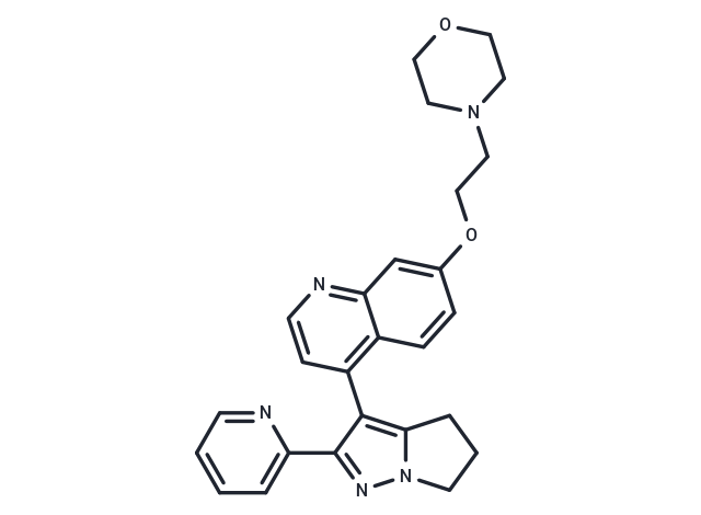
LY2109761 是一种新型选择性 TGF-β 受体 I/II 型 (TβRI/II) 双重抑制剂,Ki 分别为 38 nM 和 300 nM。


为众多的药物研发团队赋能,
让新药发现更简单!
LY2109761 是一种新型选择性 TGF-β 受体 I/II 型 (TβRI/II) 双重抑制剂,Ki 分别为 38 nM 和 300 nM。
| 规格 | 价格 | 库存 | 数量 |
|---|---|---|---|
| 1 mg | ¥ 251 | 现货 | |
| 2 mg | ¥ 367 | 现货 | |
| 5 mg | ¥ 663 | 现货 | |
| 10 mg | ¥ 1,160 | 现货 | |
| 25 mg | ¥ 2,130 | 现货 | |
| 50 mg | ¥ 3,570 | 现货 | |
| 100 mg | ¥ 5,230 | 现货 | |
| 200 mg | ¥ 7,390 | 现货 | |
| 1 mL x 10 mM (in DMSO) | ¥ 728 | 现货 |
| 产品描述 | LY2109761 is a novel selective TGF-β receptor type I/II (TβRI/II) dual inhibitor with Ki of 38 nM and 300 nM, respectively; shown to negatively affect the phosphorylation of Smad2. |
| 靶点活性 | TβRI:38 nM (Ki, cell free), TβRII:300 nM (Ki, cell free) |
| 体外活性 | 以LY2109761 (5 μM) 针对TβRI/II激酶活性几乎完全抑制了L3.6pl/GLT细胞的基础迁移率(P = 0.0107)及TGF-β1刺激下的迁移(P < 0.0001),表明L3.6pl/GLT细胞的体外迁移主要由内源性TGF-β驱动[1]。LY2109761 (0.001-0.1 μM) 显著上调E-cadherin mRNA及蛋白水平(P < 0.001),增加的E-cadherin主要定位于细胞膜,介导细胞间的锚定作用[2]。LY2109761 (10 μM) 或单独辐射 (4 Gy) 均能降低NMA-23细胞的神经球形成效率。LY2109761与辐射的联合应用在神经球形成和限制稀释实验中显示出超加性效应[3]。 |
| 体内活性 | LY2109761 (50 mg/kg, p.o.) 显著减小了肿瘤体积,并将小鼠的中位生存时间延长至45.0天,但差异并不显著。仅当LY2109761与gemcitabine联合使用时,肿瘤体积(P < 0.05)和中位生存时间显著受到影响,后者增加至77.5天(P = 0.0018) [1]。在一种原位颅内模型中,LY2109761显著减缓了肿瘤增长,延长了生存期,并延长了放射治疗引起的生存期延长。组织学分析表明,LY2109761抑制了放射促进的肿瘤侵袭,减少了肿瘤微血管密度,并减轻了间质转换 [3]。 |
| 细胞实验 | LY2109761 cytotoxicity was determined by 3 methods: the MTT assay, manual counting of viable cells, and propidium iodide staining. MTT yields a purple formazan product that is detected using a 96-well plate reader at 570 nm. Cells were plated and cultured for 2 days in a 1% fetal bovine serum medium supplemented with LY2109761 at the following concentrations: 0.001, 0.01, 0.1, 1, 10, and 20 μM. Each experimental condition was reproduced in 8 wells, and each experiment was repeated 3 times. To confirm the cytotoxic data, cells were incubated under the described conditions and stained with the vital dye trypan blue, which does not react with the cell membrane because of its negative charge. All the unstained cells were counted using a hemocytometer. Four squares were counted for each condition, and each condition was repeated in triplicate in the same experiment. Each experiment was repeated 3 times for each cell line. Bars represent the average and standard deviation of all experiments. Under the same experimental conditions, nonpermeabilized cells were stained with propidium iodide and analyzed with a flow cytometer [2]. |
| 动物实验 | Three days after the orthotopic implantation of 1.0 × 10⁶ L3.6pl/GLT tumor cells in 50 μL of HBSS, when bioluminescence imaging confirmed that tumors were well established, 40 mice were randomly allocated into four groups (n = 10 mice per group) to receive one of the following treatments. (a) Vehicle solution for 50 μL of LY2109761 twice a day p.o. (days 1–5 of each week) and 50 μL of sterile saline daily i.p. (days 2 and 5 of each week; control group). (b) LY2109761 (50 mg/kg) twice a day p.o. (days 1–5 of each week) and 50 μL of sterile saline daily i.p. (days 2 and 5 of each week). (c) Gemcitabine (25 mg/kg) daily i.p. (days 2 and 5 of each week) and p.o. vehicle for 50μL of LY2109761 twice a day (days 1–5 of each week). (d) LY2109761 (50 mg/kg) twice a day (days 1–5 of each week) and gemcitabine (25 mg/kg) daily i.p. (days 2 and 5 of each week). Treatments were continued for 4 wk. All mice were weighed weekly and observed for tumor growth. Tumor diameter was assessed with a Vernier caliper, and tumor volume (mm3) was calculated as d2 × D/2, wherein d and D represent the shortest and longest diameters, respectively. Bulky disease was considered present when the tumor burden was prominent in the mouse abdomen (tumor volume, ≥2,000 mm3). When at least 6 of 10 mice in a treatment group presented with bulky disease, the median survival duration for that group was considered to be reached. At the median survival duration of the control group, the tumor growth in mice in all groups was evaluated using the bioluminescence emitted by the tumor cells. Bioluminescence imaging was conducted using a cryogenically cooled IVIS 100 imaging system coupled to a data acquisition computer running Living Image software. The mice were sacrificed by carbon dioxide inhalation when evidence of advanced bulky disease was present. The day of sacrifice was considered the day of death for survival evaluation [1]. |
| 分子量 | 441.52 |
| 分子式 | C26H27N5O2 |
| CAS No. | 700874-71-1 |
| Smiles | C(CN1CCOCC1)Oc1ccc2c(ccnc2c1)-c1c2CCCn2nc1-c1ccccn1 |
| 密度 | 1.34 g/cm3 |
| 存储 | store at low temperature | Powder: -20°C for 3 years | In solvent: -80°C for 1 year | Shipping with blue ice. | ||||||||||||||||||||
| 溶解度信息 | DMSO: 6.88 mg/mL (15.58 mM), Sonication is recommended. Ethanol: Insoluble H2O: Insoluble | ||||||||||||||||||||
溶液配制表 | |||||||||||||||||||||
DMSO
| |||||||||||||||||||||
评论内容