购物车
- 全部删除
 您的购物车当前为空

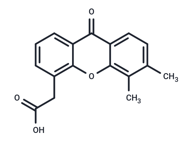
Vadimezan (DMXAA) 是一种血管破坏剂,一种鼠 STING 激动剂,也是一种细胞因子如 I 型 IFN 的诱导剂。Vadimezan 具有抗肿瘤活性,可以诱导肿瘤中的血流快速停止,但不影响正常组织中的血流。


为众多的药物研发团队赋能,
让新药发现更简单!
Vadimezan (DMXAA) 是一种血管破坏剂,一种鼠 STING 激动剂,也是一种细胞因子如 I 型 IFN 的诱导剂。Vadimezan 具有抗肿瘤活性,可以诱导肿瘤中的血流快速停止,但不影响正常组织中的血流。
| 规格 | 价格 | 库存 | 数量 |
|---|---|---|---|
| 2 mg | ¥ 415 | 现货 | |
| 5 mg | ¥ 747 | 现货 | |
| 25 mg | ¥ 2,150 | 现货 | |
| 50 mg | ¥ 3,783 | 现货 | |
| 100 mg | ¥ 4,470 | 现货 | |
| 1 mL x 10 mM (in DMSO) | ¥ 798 | 现货 |
| 产品描述 | Vadimezan (DMXAA) is a vascular disrupting agent, a murine STING agonist, and an inducer of cytokines such as type I IFN. Vadimezan has antitumor activity and induces a rapid cessation of blood flow in tumors without affecting blood flow in normal tissues. |
| 靶点活性 | DT diaphorase:20 μM(Ki) |
| 体外活性 | 方法:DLBCL 细胞 LY1 和 LY3 用 Vadimezan (0-300 µM) 处理 24 h,使用 CCK-8 assay 检测细胞活力。 结果:Vadimezan 处理以剂量依赖的方式降低 DLBCL 细胞的活力,对 LY1 和 LY3 的 IC50 分别为 177 μM 和 165 μM。[1] 方法:人肺癌细胞 A549 用 Vadimezan (0.1-1 µM) 处理 24 h,使用 Western Blot 检测靶点蛋白表达水平。 结果:Vadimezan 诱导 cytochrome c 的胞浆水平和 caspase 3 的激活显著增加,最终导致 A549 细胞凋亡死亡。[2] |
| 体内活性 | 方法:为检测体内抗肿瘤活性,将 Vadimezan (20 mg/kg) 和 BMS1166 (250 µg/mL) 腹腔注射给携带 DLBCL 肿瘤 LY1 的 Balb/c nude 小鼠,每天一次,持续八天。 结果:Vadimezan 和 BMS1166 在有效浓度下发挥作用。与单药治疗相比,联合治疗显著抑制了 GCB 样 DLBCL 细胞的生长。[1] 方法:为检测体内抗肿瘤活性,将 Vadimezan (25,5,5,25 mg/kg;25,0,0,25 mg/kg;25,25,25,25 mg/kg) 腹腔注射携带小鼠间皮瘤 AE17 的 C57BL/6J 小鼠,每三天一次,给药四次。 结果:在第 1 组中,治愈了 2/4 只小鼠,其中 2/4 只显示出长期存活,但观察到了毒性问题。在第 2 组中观察到更好、毒性更小的反应,所有 4 只小鼠均治愈并显示出长期存活。在第 3 组小鼠中,只有 1 只治愈,但没有一只长期存活,可能是由于相关的毒性问题。[3] |
| 激酶实验 | DT-diaphorase activity and kinetic analysis of enzyme inhibition : Purified DT-diaphorase enzyme activity is assayed by measuring the reduction of cytochrome c at 550 nm on a Beckman DU 650 spectrophotometer. Each assay contains cytochrome c (70 μM), NADH (variable concentrations), purified DT-diaphorase (0.032 μg), and menadione (variable concentrations) in a final volume of 1 mL Tris–HCl buffer (50 mM, pH 7.4) containing 0.14% BSA. The reaction is started by the addition of NADH. Rates of reduction are calculated over the initial part of the reaction curve (30 seconds), and results are expressed in terms of μmol cytochrome c reduced/min/mg protein using a molar extinction coefficient of 21.1 mM?1 cm?1 for reduced cytochrome c. Enzyme assays are carried out at room temperature and all reactions are performed in triplicate. Inhibition of purified DT-diaphorase activity is performed by the inclusion of DMXAA (at various concentrations) in the reaction, and inhibition characteristics are determined by varying the concentration of NADH (constant menadione) or menadione (constant NADH) at several concentrations of inhibitor. Ki values are obtained by plotting 1/V against. The activity of DT-diaphorase in DLD-1 cells is determined by measuring the dicumarol-sensitive reduction of DCPIP at 600 nm. Briefly, DLD-1 cells in mid-exponential growth are harvested by scraping into ice-cold buffer (Tris–HCl, 25 mM, pH 7.4 and 250 mM sucrose) and sonicated on ice. Enzyme assay conditions are 2 mM NADH, 40 μM DCPIP, 20 μL of dicumarol (when required) in a final volume of 1 mL Tris–HCl (25 mM, pH 7.4) containing BSA (0.7 mg/mL). Results are expressed as the dicumarol-sensitive reduction of DCPIP using a molar extinction coefficient of 21 mM?1 cm?1. Protein levels are determined using the Bradford assay |
| 细胞实验 | DLD-1 human colon carcinoma and H460 human non-small cell lung carcinoma cells are routinely maintained as monolayer cultures in RPMI 1640 culture medium supplemented with foetal calf serum (10%), sodium pyruvate (2 mM), penicillin/streptomycin (50 IU mL?1/50 μg mL-1) and l-glutamine (2 mM). Chemosensitivity is assessed using the MTT assay and all assays are performed under aerobic conditions. Briefly, cells are plated into each well of a 96-well culture plate and incubated overnight in an atmosphere containing 5% CO2. Culture medium is completely removed from each well and replaced with medium containing a range of drug concentrations. In the case of menadione alone, the duration of drug exposure is 1 hour, after which the cells are washed twice with Hanks' balanced salt solution prior to the addition of 200 μL fresh RPMI 1640 medium to each well of the plate. In the case of DMXAA alone, the duration of drug exposure is 3 hours. Following a four-day incubation, cell survival is determined using the MTT assay. For combinations of DMXAA with menadione, cells are initially set up and a non-toxic (>95% cell survival) concentration of DMXAA is selected. Cells are then initially exposed to DMXAA (2 mM) for a period of 2 hours, following which the medium is removed and replaced with medium containing the inhibitor (DMXAA at a constant concentration of 2 mM) and menadione (at a range of drug concentrations). Following a further 7-hour incubation, cells are washed twice with Hanks' balanced salt solution prior to the addition of growth medium.(Only for Reference) |
| 别名 | 伐地美生, NSC 640488, DMXAA, ASA-404, 5,6-Dimethylxanthenone-4-acetic Acid |
| 分子量 | 282.29 |
| 分子式 | C17H14O4 |
| CAS No. | 117570-53-3 |
| Smiles | Cc1ccc2c(oc3c(CC(O)=O)cccc3c2=O)c1C |
| 密度 | 1.321g/cm3 |
| 存储 | Powder: -20°C for 3 years | In solvent: -80°C for 1 year | Shipping with blue ice. | |||||||||||||||||||||||||
| 溶解度信息 | DMSO: 7.5 mg/mL (26.57 mM), Sonication is recommended. | |||||||||||||||||||||||||
| 体内实验配方 | 10% DMSO+40% PEG300+5% Tween 80+45% Saline: 0.57 mg/mL (2.02 mM), Suspension. 请按顺序添加溶剂,在添加下一种溶剂之前,尽可能使溶液澄清。如有必要,可通过加热、超声、涡旋处理进行溶解。工作液建议现配现用。以上配方仅供参考,体内配方并不是绝对的,请根据不同情况进行调整。 | |||||||||||||||||||||||||
溶液配制表 | ||||||||||||||||||||||||||
DMSO
| ||||||||||||||||||||||||||
评论内容